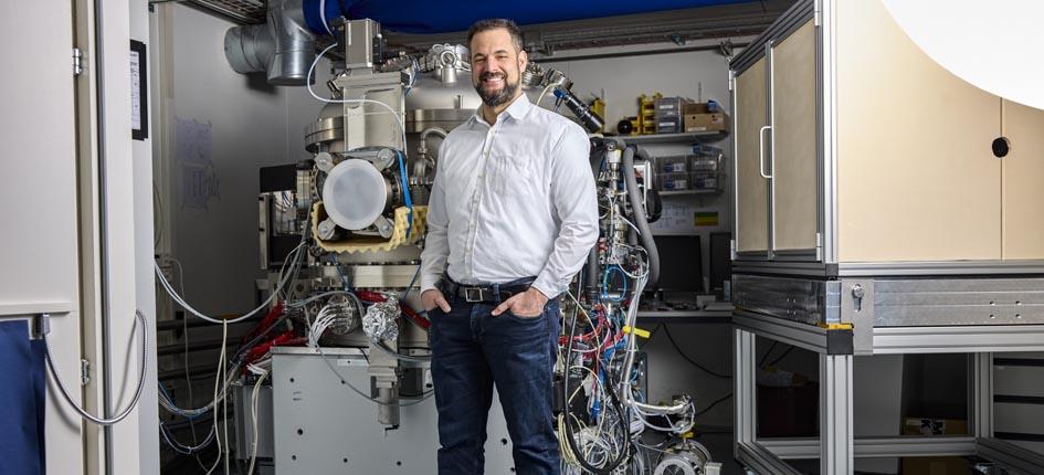
The National Institutes of Health (NIH), which is the primary agency of the United States government responsible for biomedical and public health research, has set itself the goal of decoding the architecture and wiring of the brain. In the BRAIN Initiative set up for this purpose, the NIH is teaming up with researchers from the Paul Scherrer Institute (PSI), which is based in Villigen in the canton of Aargau. PSI neurobiologist Adrian Wanner has been awarded a major grant of 2.6 million US dollars by the NIH, further details of which can be found in a press release issued by PSI. Andreas Schaefer of the Francis Crick Institute in London is also involved in the project headed up by Adrian Wanner.
“For a young research group leader to receive such a large grant, especially from another country, is by no means commonplace; it testifies to his great scientific talent and the confidence that the international community has in Switzerland as a research location”, explains Gebhard Schertler, Head of the Department of Biology and Chemistry at PSI, in the press release. “This funding will further strengthen the existing collaboration between our groups and institutes”, he concludes.
The researchers are working on a method to radically reduce the effort required to decode brain samples. To this end, they are turning to the powers of Artificial Intelligence (AI). Moreover, instead of ultra-thin cuts, wider ones in the range of 250 to 500 nanometers are used. A few nanometers are then shaved off these cuts to gradually make them thinner, with images recorded for each intermediate step. After this, a computer can generate a high-resolution 3D image from the differences between the individual images.
Over the next three years, the researchers will strive to determine whether the brain of a mouse can be completely mapped using this method. The project is being funded by the NIH over this time frame.


